BuAli Research Institute

Electron Microscope Unit

User Rating: 0 / 5

Star InactiveStar InactiveStar InactiveStar InactiveStar Inactive


MUMS Electron Microscope Unit
Buali (Avicenna) Research Institute

     
Unit Head:Dr. Alavi, Seyed Hassan 
Email: AlaviH[at]mums.ac.ir  
Phone:+98 511-7112773
staff:Mrs. Hosseini, Bibi Ashraf

Electron microscope unit is located at north-west of BouAli research institute. The unit space is about 140 square meters.
This unit has following facilities:

  • Electron microscope instrument room
  • Ultra microtome room
  • Dark room
  • Preparation of specimen room
  • Director's office


MUMS Electron Microscope Unit


History: Aim:
The transmission electron microscope is used in both metallurgy and biological sciences. It is a scientific instrument that uses a beam of electron and magnetic lenses to focus on the electron beam and magnify the image after the electrons pass through the specimen. Thus, transmission electron microscope reveals the interior of the specimen.
The specimen must be very thin and be able to withstand the high vacuum present inside the instrument. Thin sections of samples are placed on the copper grid and are stained with heavy metals. The slice of tissue can then be studied.
The EM instrument in this unit was bought in 1997  installed by Dr. Jafari.
Dr. S. Hassan Alavi has been appointed as the head of electron microscope unit since June 2006.
In addition to standard negative film
recording, the instrument is equipped with a CCD camera for digital image recording by Dr. Alavi.
 

Offer practical efficient services in the field of electron microscopy for scientists and investigators. We hope to be able to improve our services with advances in technology and update our facilities such as digital imaging as much as possible .

Lab instruments:
  • TEM. Leo910
  • Ultra microtome LKB
  • Knife maker LKB
  • Digital weighter
  • Diamond knife
  • PH meter
  • Magnetic mixer
  • Light microscope Olympus
  • Light microscope novex
  • Stereomicroscope leica
  • Oven 50

What is an Electron Microscope...

Educational activities:

In this unit, education courses on principles of transmission electron microscopy is Periodically offered for undergraduate students of Ferdowsi university, and Postgraduate students of anatomical science at MUMS.

In addition, electron microscope unit has accomplished workshops on practical training of electron microscopy many times for applicants such as faculty Members and postgraduate students.

Research activities:

Research projects in this unit have been performed with cooperation of different research Departments or independently, for example: Ultra structure study of spermatogenesis Cells after exposure of morphine in adult male mouse.

The tools and technical services for performing the thesis of postgraduate students Of MUMS and other universities have also been carried.

Clinical application of EM:

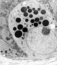
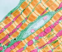
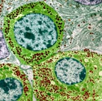
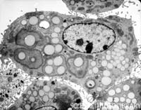
Electron microscope has been used in all areas of biological and biomedical investigations because of its ability to view the finest cell structures, and it is also used as a diagnostic tool in pathology labs for improving diagnostic and treatment of disease.

EM unit Services:
Electron microscope unit offers the tools and technical services for research and training for those who do not necessarily want to operate the electron microscope instrument, or to prepare specimen themselves. The staff of the Electron microscope unit offer training and practical services including Microscope operation, specimen preparation, digital photography and Image analysis.

New services in  IMU:
 Electron microscpe unit offers technical services in Immunoelectron microscopy to detect the intracellular location in structures of particular proteins.
 
 
Specimen Preparation for TEM:
 
Procedure:

1. Primary fixation (glutaraldehyde 2-4%)

2. Secondary fixation (osmium tetroxide 1-2%)

3. Dehydration

4. Infiltration

5. Embedding

6. Sectioning

7. Staining

MUMS Electron Microscope Unit